Background: Small cell lung cancer (SCLC) is an aggressive neuroendocrine malignancy characterized by early, widespread hematogenous dissemination. Standard metastatic sites include the liver, brain, bones, and adrenal glands. Solitary skeletal muscle metastasis (SMM) as the initial presenting feature of occult SCLC is exceedingly rare, often leading to diagnostic delay or misdiagnosis as a soft-tissue sarcoma or hematoma.
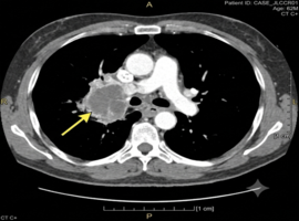
Case Presentation: A 62-year-old male with a history of heavy tobacco use presented with a 3-week history of a progressively painful, firm swelling in his left thigh, without overt pulmonary symptoms. Soft-tissue ultrasound and magnetic resonance imaging (MRI) revealed a deeply seated, hypervascular intramuscular mass within the vastus lateralis. Histopathological evaluation via core-needle biopsy demonstrated sheets of small, round blue cells with crushed chromatin, high mitotic activity, and nuclear molding. Immunohistochemistry was strongly positive for synaptophysin, chromogranin A, and TTF-1, confirming high-grade neuroendocrine carcinoma consistent with metastasized SCLC. Subsequent contrast-enhanced thoracic CT identified a 2.8 cm primary central hilar mass with mediastinal lymphadenopathy. The patient was staged with extensive-stage SCLC (ES-SCLC) and initiated on systemic platinum-based chemotherapy combined with immune checkpoint inhibition, resulting in significant regression of both the primary lesion and the thigh mass.
Conclusion: Solitary intramuscular metastasis should be considered in the differential diagnosis of soft-tissue masses in older patients, particularly those with a significant smoking history. Biopsy with immunohistochemical staining is crucial to differentiate SMM from primary soft-tissue neoplasms and prevent inappropriate surgical interventions.
Keywords: Solitary Skeletal Muscle; Metastasis; Small Cell Lung Cancer; Thoracic Oncology